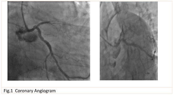
Firstly, we have perform an mid LAD disocclusion with a 3x18mm Everolimus drug eluting stent.
After an Optical coherence tomography imaging (C7-XR TM OCT Intravascular Imaging System, St. Jude Medical) was planned, and it was shown a ruptured plaque with a cavity formed inside it, corresponding to the ambigious image at angiogram (fig.2).


After quantitative measure of the reference diameter and reference area of the LM (4.4mm) and the length of the lesion by OCT, PCI of LM to LAD was performed with 4 x 24 mm
Everolimus eluting stent implantation, using the technique of POT-Side-RePOT ( 4.5 x 10mm Balloon). Next the second OCT run was performed after stent implantation and it have shown a good apposition of the stent struts to the vessel wall, and an adequate stent expansion (fig.3)


Discussion
Intravascular imaging (IVUS or OCT) allows real-time tomographic assessment of vessel size, lumen area, and plaque composition and volume.
OCT is a light-based modality of intravascular imaging with higher axial resolution compared with IVUS (15 vs 150 mm), but it requires complete blood clearance from the lumen for imaging and that it has more limited penetration.
In angiographically hazy lesions, OCT often detects ruptured plaques with a thrombus attached to the site of rupture of the fibrous caps over a partially emptied lipid pool 1 . The ability of OCT to address luminal areas and identify underexpansion, malapposition, uneven
stent strut distribution, or small intra-stent thrombotic formations makes the technique a very attractive tool for the prevention of thrombosis 2 .
DOCTORS Study has shown that in patients with NSTE-ACS, OCT provided useful additional information beyond that obtained by angiography alone and was associated with better functional outcome as assessed by FFR 3 .
1. Prati F, et al. for the Expert’s OCT Review Document. Expert review document on methodology and clinical applications of OCT. Physical principles, methodology of image acquisition and clinical application for assessment of coronary arteries and atherosclerosis. Eur Heart J 2010;31:401–415.
2. Prati, F et al. Expert review document part 2: methodology, terminology and clinical applications of optical coherence tomography for the assessment of interventional procedures. European Heart Journal, 33(20), 2513–2520. doi:10.1093/eurheartj/ehs095
3. Meneveau, N et al. Optical Coherence Tomography to Optimize Results of Percutaneous Coronary Intervention in Patients with Non–ST-Elevation Acute Coronary SyndromeClinical Perspective.
Circulation, 134(13), 906–917. doi:10.1161/circulationaha.116.024393